Copyright
©2014 Baishideng Publishing Group Inc.
World J Gastroenterol. Oct 28, 2014; 20(40): 14760-14777
Published online Oct 28, 2014. doi: 10.3748/wjg.v20.i40.14760
Published online Oct 28, 2014. doi: 10.3748/wjg.v20.i40.14760
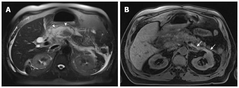
Figure 5 Acute interstitial edematous pancreatitis with acute peripancreatic fluid collection.
A: Axial single-shot turbo spin-echo T2-weighted (HASTE) image with fat-suppression; B: Axial 3D-GRE T1-weighted image with fat-suppression. The pancreas shows mild lace-like increased T2 signal involving the pancreatic parenchyma (A), with minimally decreased T1 signal intensity and mild peripancreatic stranding (B), associated with peripancreatic fluid collections (arrowheads, A) and small proteinaceous fluid at the left anterior para- and peri-renal spaces (arrows); both associated with imperceptible wall in keeping with acute interstitial edematous pancreatitis and acute peripancreatic fluid collection. Minimal ascites is also seen (A).
- Citation: Manikkavasakar S, AlObaidy M, Busireddy KK, Ramalho M, Nilmini V, Alagiyawanna M, Semelka RC. Magnetic resonance imaging of pancreatitis: An update. World J Gastroenterol 2014; 20(40): 14760-14777
- URL: https://www.wjgnet.com/1007-9327/full/v20/i40/14760.htm
- DOI: https://dx.doi.org/10.3748/wjg.v20.i40.14760


