Copyright
©2014 Baishideng Publishing Group Inc.
World J Gastroenterol. Oct 28, 2014; 20(40): 14747-14759
Published online Oct 28, 2014. doi: 10.3748/wjg.v20.i40.14747
Published online Oct 28, 2014. doi: 10.3748/wjg.v20.i40.14747
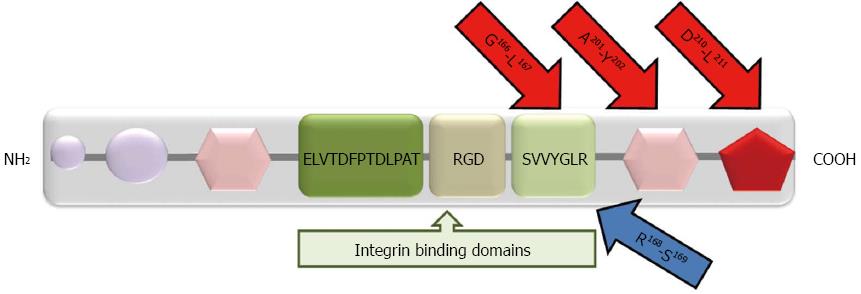
Figure 2 Structural domains of osteopontin.
Purple circles: Matrix binding domains; pink hexagons: Calcium binding sites; Red pentagon: Heparin binding site. There are three integrin binding sequences: (1) Arginine-glycine-aspartic acid (RGD); (2) Serine-valine-valine-tyrosine-glutamate-leucine-arginine (SVVYGLR); and (3) ELVTDFPTDLPAT. MMP cleavage sites (G166-L167; A201-Y202; D210-L211) are shown by red arrows. The thrombin cleavage site (R168-S169) is shown by blue arrow.
- Citation: Kaleağasıoğlu F, Berger MR. SIBLINGs and SPARC families: Their emerging roles in pancreatic cancer. World J Gastroenterol 2014; 20(40): 14747-14759
- URL: https://www.wjgnet.com/1007-9327/full/v20/i40/14747.htm
- DOI: https://dx.doi.org/10.3748/wjg.v20.i40.14747


