Copyright
©2014 Baishideng Publishing Group Co.
World J Gastroenterol. Jan 28, 2014; 20(4): 1123-1126
Published online Jan 28, 2014. doi: 10.3748/wjg.v20.i4.1123
Published online Jan 28, 2014. doi: 10.3748/wjg.v20.i4.1123
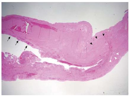
Figure 4 Pathological findings of resected specimen showing duodenal wall with partially denuded epithelium (hematoxylin and eosin staining, ×12.
5). The mucosal lining (black arrows), smooth muscle coat (black arrowheads), and glands (white arrowheads) were noticed.
- Citation: Byun J, Oh HM, Kim SH, Kim HY, Jung SE, Park KW, Kim WS. Laparoscopic partial cystectomy with mucosal stripping of extraluminal duodenal duplication cysts. World J Gastroenterol 2014; 20(4): 1123-1126
- URL: https://www.wjgnet.com/1007-9327/full/v20/i4/1123.htm
- DOI: https://dx.doi.org/10.3748/wjg.v20.i4.1123


