Copyright
©2014 Baishideng Publishing Group Inc.
World J Gastroenterol. Oct 14, 2014; 20(38): 14076-14078
Published online Oct 14, 2014. doi: 10.3748/wjg.v20.i38.14076
Published online Oct 14, 2014. doi: 10.3748/wjg.v20.i38.14076
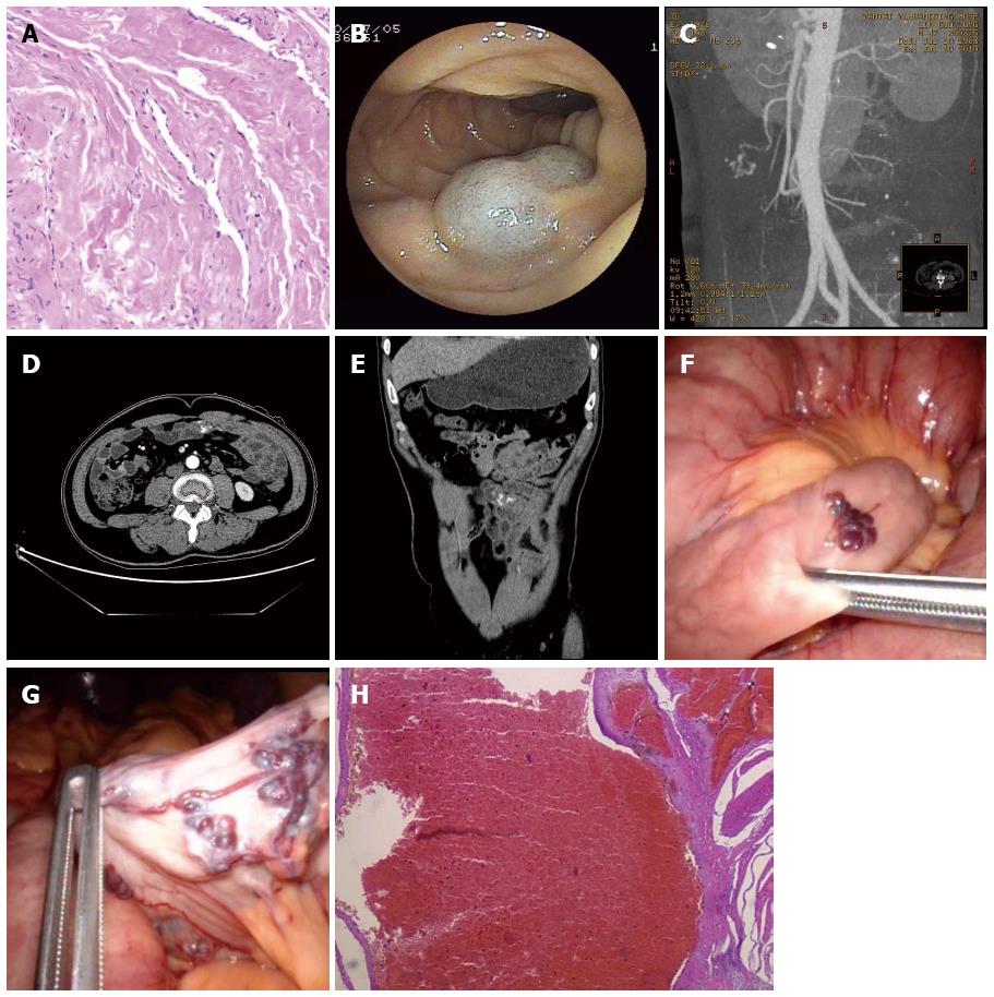
Figure 1 Skin, enteroscopy, abdominal vascular enhanced computed tomography, laparoscopy and pathological findings of the patient.
A: Pathological analysis showed cavernous angioma on the right knee (× 400); B: Angioma was indicated by oral approach enteroscopy; C: Arterior-venous malformation at the branch of the superior mesenteric artery was shown by abdominal vascular enhanced computed tomography (CT); D: Contrast media remained in the arterior-venous malformation, as shown by abdominal vascular enhanced CT at the cross section; E: Contrast media remained in the arterior-venous malformation, as shown by abdominal vascular enhanced CT in a reconstruction of a coronal view; F: A small intestinal angioma was detected by a laparoscopic approach; the tissue in this image corresponds to B; G: Arterior-venous malformation was found by a laparoscopic approach; the tissue in this image corresponds to C-E; H: By pathological examination, the resected small intestinal arterior-venous malformation was shown to have a vessel wall that was irregular, distorted and dilated (× 400).
- Citation: Cui J, Huang LY, Lin SJ, Yi LZ, Wu CR, Zhang B. Small intestinal vascular malformation bleeding: A case report with imaging findings. World J Gastroenterol 2014; 20(38): 14076-14078
- URL: https://www.wjgnet.com/1007-9327/full/v20/i38/14076.htm
- DOI: https://dx.doi.org/10.3748/wjg.v20.i38.14076


