Copyright
©2014 Baishideng Publishing Group Inc.
World J Gastroenterol. Oct 14, 2014; 20(38): 13956-13965
Published online Oct 14, 2014. doi: 10.3748/wjg.v20.i38.13956
Published online Oct 14, 2014. doi: 10.3748/wjg.v20.i38.13956
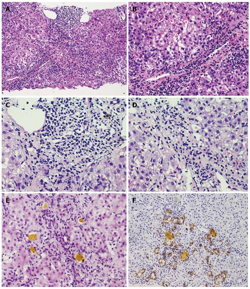
Figure 3 Nonsteroidal anti-inflammatory drugs-induced acute hepatitis.
A: There is a bridging necrosis zone around the central vein (HE, × 100); B: Vacuole and necrosis are found in hepatic cells around the necrosis zone (HE, × 400); C: A substantial inflammatory cells including lymphocytes, plasma cells, and eosinophils infiltrate can be seen in the portal tract (HE, × 400); D: Hepatic necrosis and lymphocytic infiltration, including some eosinophils (arrow), in the collapsed parenchyma can be observed (HE, × 400); E: NSAID-induced acute cholestasis; E: Small bile duct dilation and cholestasis can be seen in the portal area (HE, × 400); F: The cholestasis accompanying hepatocellular vacuolation is presented (HE, × 100). HE: Hematoxylin and eosin staining; NSAID: Nonsteroidal anti-inflammatory drug.
- Citation: Cao YL, Tian ZG, Wang F, Li WG, Cheng DY, Yang YF, Gao HM. Characteristics and clinical outcome of nonsteroidal anti-inflammatory drug-induced acute hepato-nephrotoxicity among Chinese patients. World J Gastroenterol 2014; 20(38): 13956-13965
- URL: https://www.wjgnet.com/1007-9327/full/v20/i38/13956.htm
- DOI: https://dx.doi.org/10.3748/wjg.v20.i38.13956


