Copyright
©2014 Baishideng Publishing Group Inc.
World J Gastroenterol. Oct 14, 2014; 20(38): 13956-13965
Published online Oct 14, 2014. doi: 10.3748/wjg.v20.i38.13956
Published online Oct 14, 2014. doi: 10.3748/wjg.v20.i38.13956
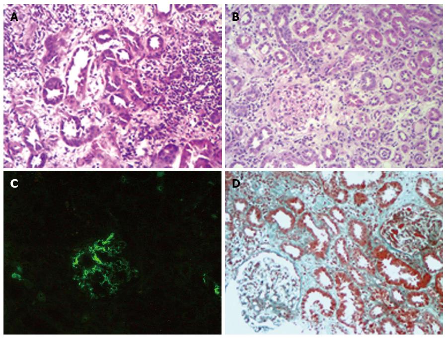
Figure 2 Nonsteroidal anti-inflammatory drugs-induced acute interstitial nephritis and acute tubulointerstitial disease.
A: Substantial inflammatory cells, including lymphocytes and eosinophils infiltrate, can be seen in the renal interstitium. The tubular epithelium undergoes necrosis, which can be seen by denudation of tubular epithelial cells with the rupturing of the tubular basement membrane (HE, × 200); B: Edema and inflammatory infiltrate in the interstitium cause the renal tubules to be abnormally widely spaced with no changes to the glomerulus. The infiltrate includes inflammatory cells (eosinophils and lymphocytes), and there is obvious disorder of the renal tubular epithelium with significant edema (HE, × 200); C: Presence of IgA is seen throughout the mesangium by immunofluorescence (Fluorescein isothiocyanate anti-IgA, × 200); D: Proliferation of mesangial cells is seen along with focal inflammation of glomeruli. The denudation of the tubular epithelial cells, the edema and inflammatory infiltration in the interstitum are indicted (Masson staining, × 200). HE: Hematoxylin and eosin staining.
- Citation: Cao YL, Tian ZG, Wang F, Li WG, Cheng DY, Yang YF, Gao HM. Characteristics and clinical outcome of nonsteroidal anti-inflammatory drug-induced acute hepato-nephrotoxicity among Chinese patients. World J Gastroenterol 2014; 20(38): 13956-13965
- URL: https://www.wjgnet.com/1007-9327/full/v20/i38/13956.htm
- DOI: https://dx.doi.org/10.3748/wjg.v20.i38.13956


