Copyright
©2014 Baishideng Publishing Group Inc.
World J Gastroenterol. Oct 14, 2014; 20(38): 13842-13862
Published online Oct 14, 2014. doi: 10.3748/wjg.v20.i38.13842
Published online Oct 14, 2014. doi: 10.3748/wjg.v20.i38.13842
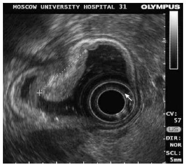
Figure 6 Same patient.
Endosonography of the lesion 0-IIa + Is. Area of the lesion (25 mm in size): Thickening of mucosa up to 5-7 mm; submucosal layer is clear under the tumor; lymph nodes are not visualized.
- Citation: Pasechnikov V, Chukov S, Fedorov E, Kikuste I, Leja M. Gastric cancer: Prevention, screening and early diagnosis. World J Gastroenterol 2014; 20(38): 13842-13862
- URL: https://www.wjgnet.com/1007-9327/full/v20/i38/13842.htm
- DOI: https://dx.doi.org/10.3748/wjg.v20.i38.13842


