Copyright
©2014 Baishideng Publishing Group Inc.
World J Gastroenterol. Oct 14, 2014; 20(38): 13842-13862
Published online Oct 14, 2014. doi: 10.3748/wjg.v20.i38.13842
Published online Oct 14, 2014. doi: 10.3748/wjg.v20.i38.13842
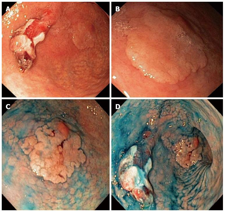
Figure 2 Esophagogastroduodenoscopy of the same patient.
A: The flat lesion in the background can been viewed more easily when better lit; B: Closer view of the superficial elevated lesion; C: After chromoendoscopy with indigo carmine - a roundish lesion 25 mm in diameter can be seen, with a smooth lobulated surface and a 6-mm, reddish protrusion in the distal part; type 0-IIa+Is according to Paris classification; D: Due to the marked inflammation and presence of intestinal metaplasia the precise proximal margin of the lesion is still unclear, even with the use of chromoendscopy.
- Citation: Pasechnikov V, Chukov S, Fedorov E, Kikuste I, Leja M. Gastric cancer: Prevention, screening and early diagnosis. World J Gastroenterol 2014; 20(38): 13842-13862
- URL: https://www.wjgnet.com/1007-9327/full/v20/i38/13842.htm
- DOI: https://dx.doi.org/10.3748/wjg.v20.i38.13842


