Copyright
©2014 Baishideng Publishing Group Inc.
World J Gastroenterol. Oct 7, 2014; 20(37): 13625-13631
Published online Oct 7, 2014. doi: 10.3748/wjg.v20.i37.13625
Published online Oct 7, 2014. doi: 10.3748/wjg.v20.i37.13625
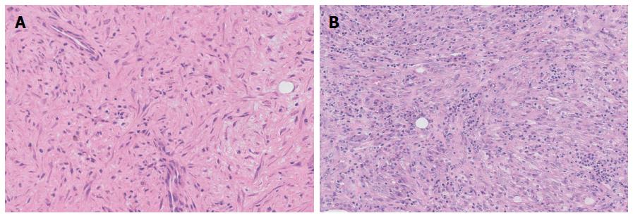
Figure 3 Pathological images of two patients.
A: Histological examination showing a proliferation of myofibroblasts and infiltration of inflammation cells (HE stain, 200 ×); B: Histological examination showing a proliferation of spindled cells, likely myofibroblasts, mixed with abundant lymphocytes and plasma cells (HE stain, 200 ×).
- Citation: Zhao JJ, Ling JQ, Fang Y, Gao XD, Shu P, Shen KT, Qin J, Sun YH, Qin XY. Intra-abdominal inflammatory myofibroblastic tumor: Spontaneous regression. World J Gastroenterol 2014; 20(37): 13625-13631
- URL: https://www.wjgnet.com/1007-9327/full/v20/i37/13625.htm
- DOI: https://dx.doi.org/10.3748/wjg.v20.i37.13625


