Copyright
©2014 Baishideng Publishing Group Inc.
World J Gastroenterol. Oct 7, 2014; 20(37): 13625-13631
Published online Oct 7, 2014. doi: 10.3748/wjg.v20.i37.13625
Published online Oct 7, 2014. doi: 10.3748/wjg.v20.i37.13625
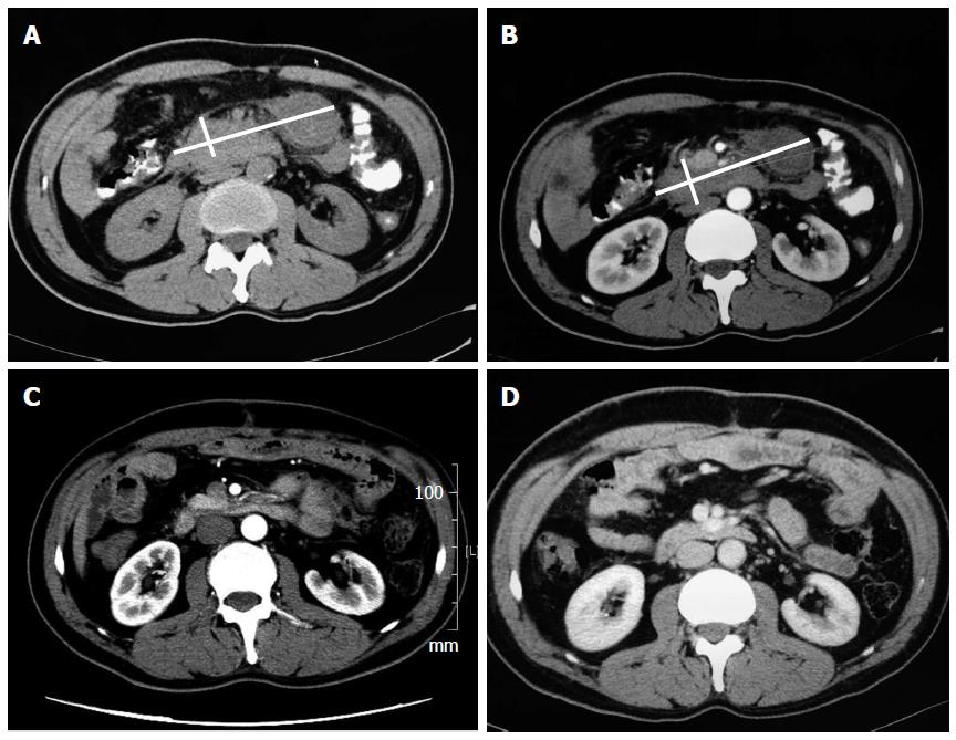
Figure 1 Computed tomography imaging changes of patient No.
1 A: A plain computed tomography (CT) scan on admission showing a large nodular mass (white lines mark the maximum and minimum diameter) located in the upper quadrant of the peritoneum and encasing the surrounding soft tissues; B: An enhancement CT scan showing the mass to be multinodular. Some parts of the mass were mildly enhanced, while others were cystic degenerated; C: A CT scan obtained 3 wk after the operation identified the spontaneous regression of the tumor; D: A CT scan obtained 3 mo later did not show any indication of relapse.
- Citation: Zhao JJ, Ling JQ, Fang Y, Gao XD, Shu P, Shen KT, Qin J, Sun YH, Qin XY. Intra-abdominal inflammatory myofibroblastic tumor: Spontaneous regression. World J Gastroenterol 2014; 20(37): 13625-13631
- URL: https://www.wjgnet.com/1007-9327/full/v20/i37/13625.htm
- DOI: https://dx.doi.org/10.3748/wjg.v20.i37.13625


