Copyright
©2014 Baishideng Publishing Group Inc.
World J Gastroenterol. Oct 7, 2014; 20(37): 13615-13619
Published online Oct 7, 2014. doi: 10.3748/wjg.v20.i37.13615
Published online Oct 7, 2014. doi: 10.3748/wjg.v20.i37.13615
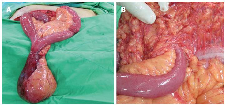
Figure 3 Intraoperative photographs.
A: The herniated Meckel’s diverticulum and small bowel, showing the completion of the manual reduction of the mesenteric volvulus; B: The inlet of the internal hernia in the transverse mesocolon.
- Citation: Wu SY, Ho MH, Hsu SD. Meckel's diverticulum incarcerated in a transmesocolic internal hernia. World J Gastroenterol 2014; 20(37): 13615-13619
- URL: https://www.wjgnet.com/1007-9327/full/v20/i37/13615.htm
- DOI: https://dx.doi.org/10.3748/wjg.v20.i37.13615


