Copyright
©2014 Baishideng Publishing Group Inc.
World J Gastroenterol. Oct 7, 2014; 20(37): 13538-13545
Published online Oct 7, 2014. doi: 10.3748/wjg.v20.i37.13538
Published online Oct 7, 2014. doi: 10.3748/wjg.v20.i37.13538
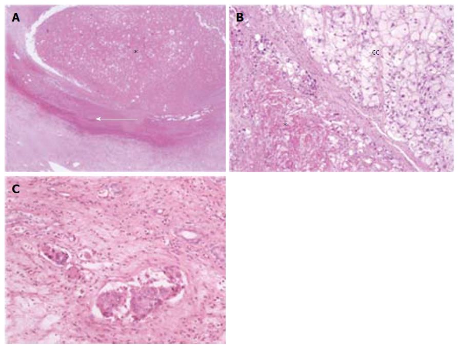
Figure 1 Different histological pictures after transarterial chemoembolization.
A: A case of totally necrotic nodule (asterisk), with a well-visible fibrotic capsule (white arrow); B: A case of clear cell (CC) hepatocellular carcinoma with only partial necrosis (asterisk); C: Amicrovascular invasion. Haematoxylin-Eosin stain; Magnification × 2 (A) and × 10 (B, C).
- Citation: Vasuri F, Malvi D, Rosini F, Baldin P, Fiorentino M, Paccapelo A, Ercolani G, Pinna AD, Golfieri R, Morselli-Labate AM, Grigioni WF, D’Errico-Grigioni A. Revisiting the role of pathological analysis in transarterial chemoembolization-treated hepatocellular carcinoma after transplantation. World J Gastroenterol 2014; 20(37): 13538-13545
- URL: https://www.wjgnet.com/1007-9327/full/v20/i37/13538.htm
- DOI: https://dx.doi.org/10.3748/wjg.v20.i37.13538


