Copyright
©2014 Baishideng Publishing Group Inc.
World J Gastroenterol. Oct 7, 2014; 20(37): 13453-13465
Published online Oct 7, 2014. doi: 10.3748/wjg.v20.i37.13453
Published online Oct 7, 2014. doi: 10.3748/wjg.v20.i37.13453
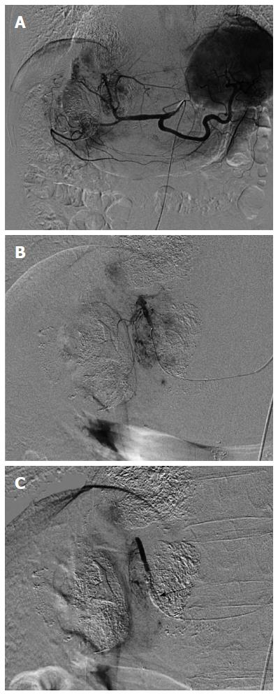
Figure 1 Selective arteriography with and without balloon-occluded feeding artery in a 72 year-old man with multiple hepatocellular carcinomas.
A: Celiac arteriography revealed multiple hepatocellular carcinomas (HCCs) in the liver; B: Opacified HCCs were shown by medial segmental arteriography without balloon-occluded feeding artery; C: In contrast, the arteriography with balloon occlusion showed faint opacification of HCCs and nontumoral liver parenchyma was opacified. Arrow indicated an inflated microballoon.
- Citation: Murata S, Mine T, Sugihara F, Yasui D, Yamaguchi H, Ueda T, Onozawa S, Kumita SI. Interventional treatment for unresectable hepatocellular carcinoma. World J Gastroenterol 2014; 20(37): 13453-13465
- URL: https://www.wjgnet.com/1007-9327/full/v20/i37/13453.htm
- DOI: https://dx.doi.org/10.3748/wjg.v20.i37.13453


