Copyright
©2014 Baishideng Publishing Group Inc.
World J Gastroenterol. Sep 28, 2014; 20(36): 13195-13199
Published online Sep 28, 2014. doi: 10.3748/wjg.v20.i36.13195
Published online Sep 28, 2014. doi: 10.3748/wjg.v20.i36.13195
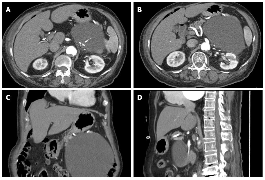
Figure 2 Computed tomography scans show a large low-attenuation lesion measuring 16 cm in the pancreatic tail with luminal calcification (white arrow) (A), the cyst abutting the pancreas (B), a large cystic mass in the retroperitoneum (C); the cystic mass below the pancreas (D).
- Citation: Jung HI, Ahn T, Son MW, Kim Z, Bae SH, Lee MS, Kim CH, Cho HD. Adrenal lymphangioma masquerading as a pancreatic tail cyst. World J Gastroenterol 2014; 20(36): 13195-13199
- URL: https://www.wjgnet.com/1007-9327/full/v20/i36/13195.htm
- DOI: https://dx.doi.org/10.3748/wjg.v20.i36.13195


