Copyright
©2014 Baishideng Publishing Group Inc.
World J Gastroenterol. Sep 28, 2014; 20(36): 12993-13005
Published online Sep 28, 2014. doi: 10.3748/wjg.v20.i36.12993
Published online Sep 28, 2014. doi: 10.3748/wjg.v20.i36.12993
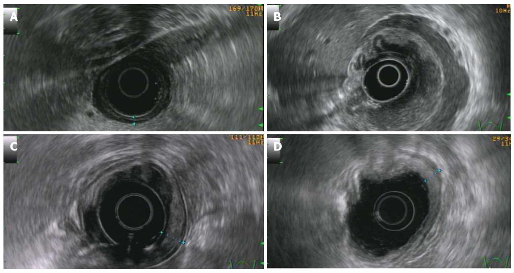
Figure 3 Endoscopic ultrasonography (radial scanning).
A: Disruption of the second layer with preservation of the other layers and no nodal involvement (T1 N0); B: Diffuse infiltrative pattern: diffuse trans-mural involvement of the gastric wall with irregular layer border and not uniform echogenicity. Diffuse circumferential tickening of the second, third and fourth layers; the is still preserved (T2 N0); C: Localized hypoechoic alteration limited at the third layer, whit a mild thickening of the stomach wall and circumscribed to a small portion of the gastric circumference (T1 sm N0); D: Persistent tickening of the first and second layer in a patient affected by diffuse large B cell lymphoma in complete remission after the immuno-chemotherapy performed 2 years before.
- Citation: Vetro C, Romano A, Amico I, Conticello C, Motta G, Figuera A, Chiarenza A, Raimondo CD, Giulietti G, Bonanno G, Palumbo GA, Raimondo FD. Endoscopic features of gastro-intestinal lymphomas: From diagnosis to follow-up. World J Gastroenterol 2014; 20(36): 12993-13005
- URL: https://www.wjgnet.com/1007-9327/full/v20/i36/12993.htm
- DOI: https://dx.doi.org/10.3748/wjg.v20.i36.12993


