Copyright
©2014 Baishideng Publishing Group Inc.
World J Gastroenterol. Sep 21, 2014; 20(35): 12696-12700
Published online Sep 21, 2014. doi: 10.3748/wjg.v20.i35.12696
Published online Sep 21, 2014. doi: 10.3748/wjg.v20.i35.12696
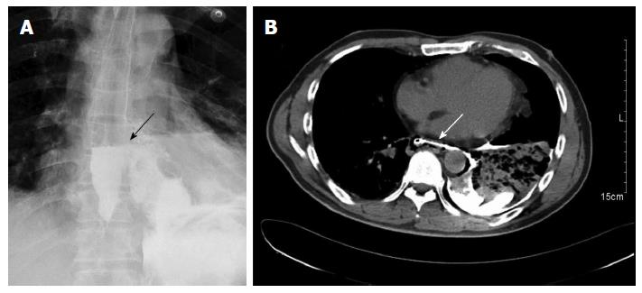
Figure 1 A gastrografin swallow, showing extravasation of contrast in the left chest (arrow) (A), computed tomography of the chest with oral contrast, demonstrating a lower left esophageal tear with mediastinal leakage of oral contrast medium (arrow) and a left pleural effusion (B).
- Citation: Shen G, Chai Y, Zhang GF. Successful surgical strategy in a late case of Boerhaave’s syndrome. World J Gastroenterol 2014; 20(35): 12696-12700
- URL: https://www.wjgnet.com/1007-9327/full/v20/i35/12696.htm
- DOI: https://dx.doi.org/10.3748/wjg.v20.i35.12696


