Copyright
©2014 Baishideng Publishing Group Inc.
World J Gastroenterol. Sep 21, 2014; 20(35): 12682-12686
Published online Sep 21, 2014. doi: 10.3748/wjg.v20.i35.12682
Published online Sep 21, 2014. doi: 10.3748/wjg.v20.i35.12682
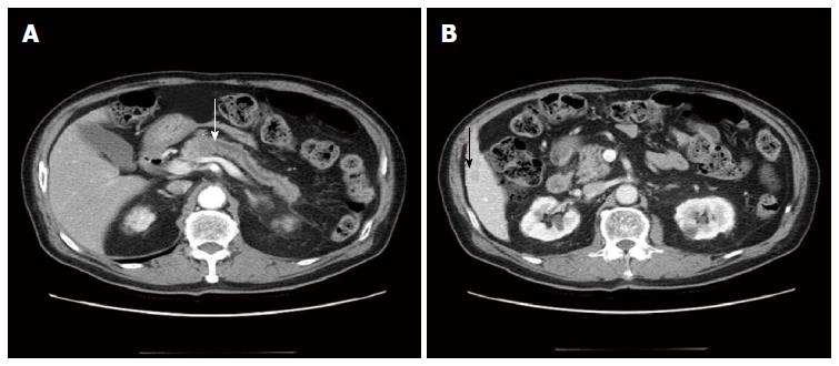
Figure 1 Axial pancreas-enhanced computed tomography findings.
A: A 2.2 cm low attenuated mass in the pancreas body with diffuse pancreas duct dilatation (white arrow: pancreatic head mass); B: A 1.3 cm low attenuated hepatic nodule in S5 (black arrow: hepatic mass).
- Citation: Kim HS, Kim JI, Jeong M, Seo JH, Kim IK, Cheung DY, Kim TJ, Kang CS. Pancreatic adenocarcinosarcoma of monoclonal origin: A case report. World J Gastroenterol 2014; 20(35): 12682-12686
- URL: https://www.wjgnet.com/1007-9327/full/v20/i35/12682.htm
- DOI: https://dx.doi.org/10.3748/wjg.v20.i35.12682


