Copyright
©2014 Baishideng Publishing Group Inc.
World J Gastroenterol. Sep 21, 2014; 20(35): 12678-12681
Published online Sep 21, 2014. doi: 10.3748/wjg.v20.i35.12678
Published online Sep 21, 2014. doi: 10.3748/wjg.v20.i35.12678
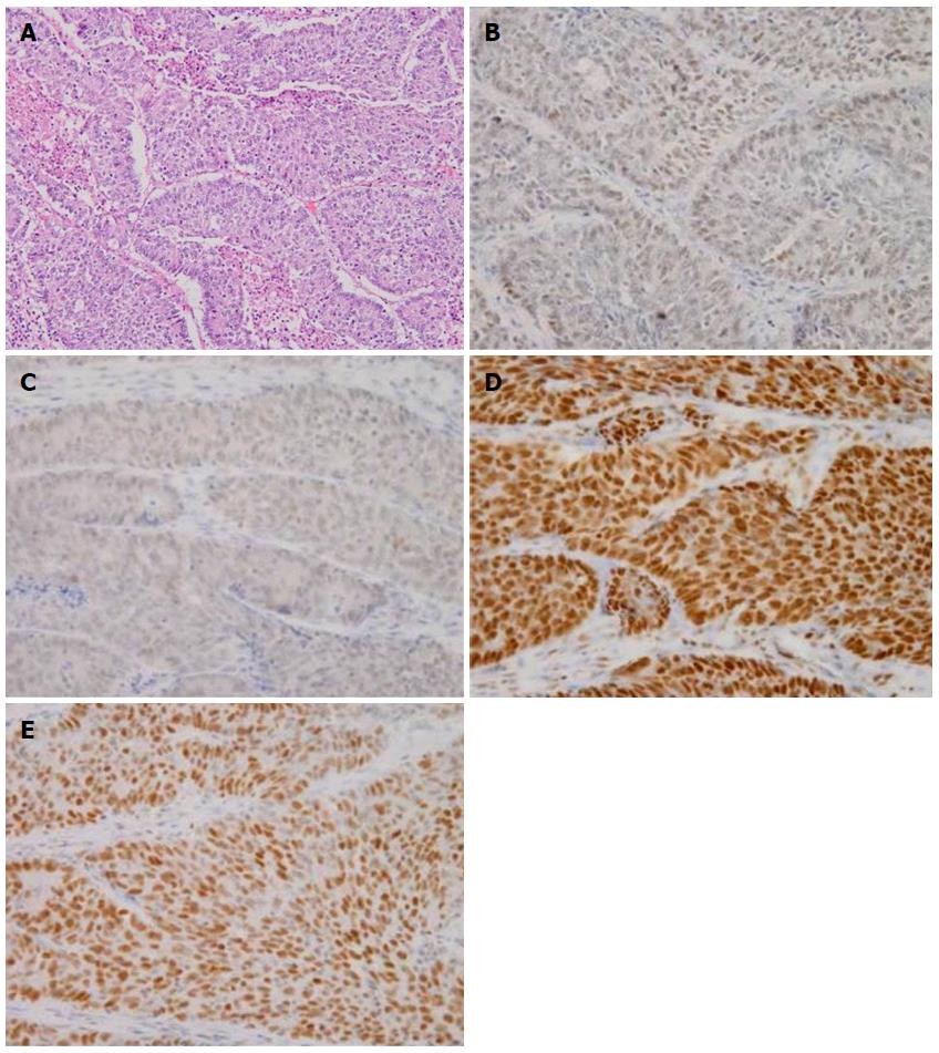
Figure 4 Microscopic findings and immunohistochemistry for deoxyribonucleic acid mismatch repaire proteins (× 200).
The tumor is a poorly differentiated adenocarcinoma. Immunohistochemical expression of mutL homolog 1 (MLH1), postmeiotic segregation increased 2 (PMS2), mutS homologue 2 (MSH2) and mutS homologue 6 (MSH6) is retained. HE: Hematoxylin and eosin stain. A: HE; B: MLH1, C: PMS2; D: MSH2, E: MSH6.
- Citation: Inada R, Nagasaka T, Toshima T, Mori Y, Kondo Y, Kishimoto H, Fujiwara T. Intussusception due to rectal adenocarcinoma in a young adult: A case report. World J Gastroenterol 2014; 20(35): 12678-12681
- URL: https://www.wjgnet.com/1007-9327/full/v20/i35/12678.htm
- DOI: https://dx.doi.org/10.3748/wjg.v20.i35.12678


