Copyright
©2014 Baishideng Publishing Group Inc.
World J Gastroenterol. Sep 21, 2014; 20(35): 12673-12677
Published online Sep 21, 2014. doi: 10.3748/wjg.v20.i35.12673
Published online Sep 21, 2014. doi: 10.3748/wjg.v20.i35.12673
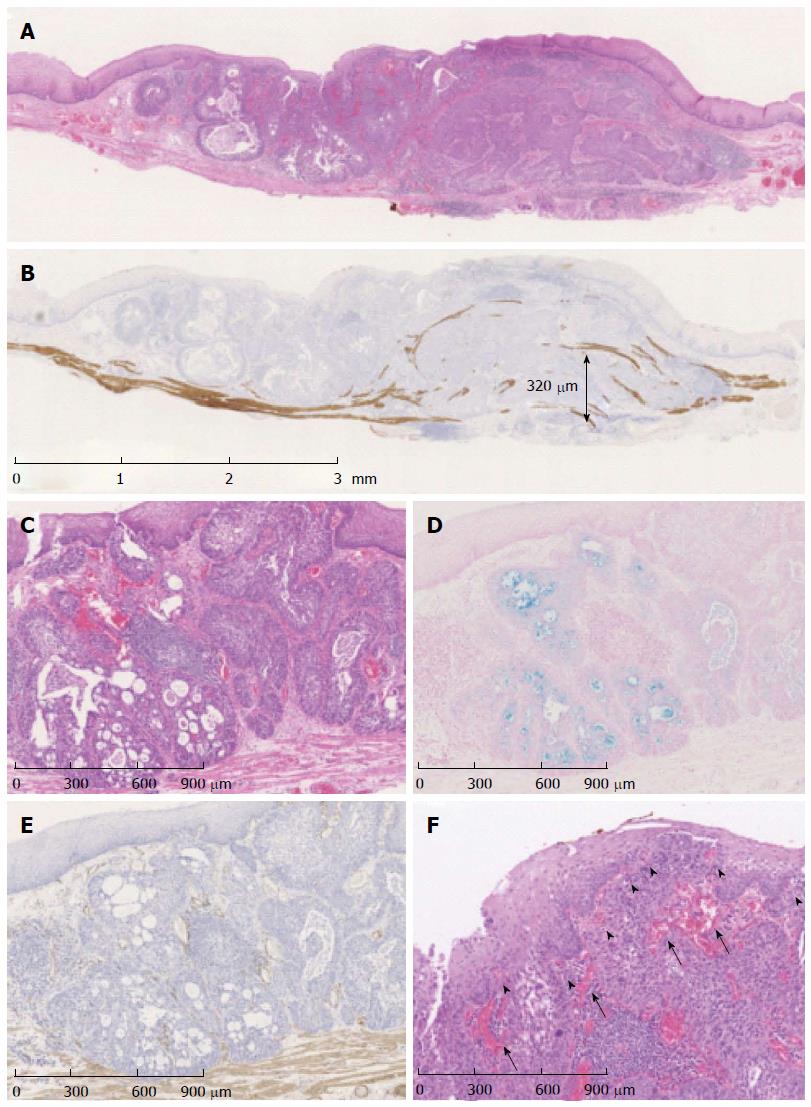
Figure 5 Hyperchromatic tumor cells.
A: Histologically, basal cell-like hyperchromatic tumor cells proliferated mainly in the lamina propria; B: Immunostaining with desmin suggested that the tumor invaded the submucosa at a depth of 320 μm; C: The tumor cells formed solid nests and microcystic structures; D: The microcystic structures contained an Alcian blue-positive mucoid matrix; E: Immunostaining for α-SMA was negative; F: Thin vessels (arrowheads) were observed in the intra-epithelial papilla, and thick vessels (arrows) were observed around the solid nests just below the epithelium.
- Citation: Kai Y, Kato M, Hayashi Y, Akasaka T, Shinzaki S, Nishida T, Tsujii M, Morii E, Takehara T. Esophageal early basaloid squamous carcinoma with unusual narrowband imaging magnified endoscopy findings. World J Gastroenterol 2014; 20(35): 12673-12677
- URL: https://www.wjgnet.com/1007-9327/full/v20/i35/12673.htm
- DOI: https://dx.doi.org/10.3748/wjg.v20.i35.12673


