Copyright
©2014 Baishideng Publishing Group Inc.
World J Gastroenterol. Sep 21, 2014; 20(35): 12628-12636
Published online Sep 21, 2014. doi: 10.3748/wjg.v20.i35.12628
Published online Sep 21, 2014. doi: 10.3748/wjg.v20.i35.12628
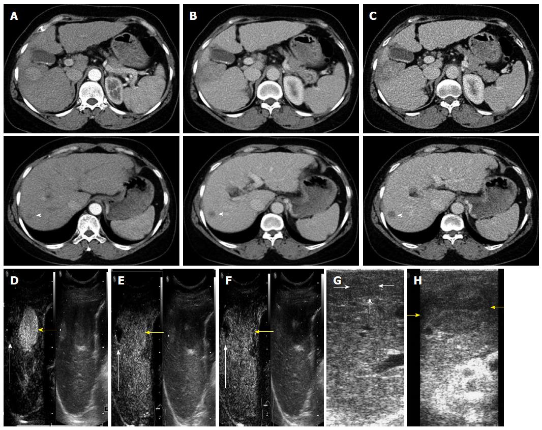
Figure 2 A 55-year-old woman underwent right hepatic partial resection.
A-C: both lesions, displayed in the arterial, portal venous and equilibrium phases, were diagnosed as HCC by CT (white arrow); D-F: the HCC (yellow arrow) displayed wash-in in arterial phase and wash-out in late phases via CEUS; another lesion (white arrow), diagnosed as necrosis by final histopathology, displayed iso-enhancement in the three phases; G: the HCC identified by IOUS; H: the necrosis also falsely diagnosed as HCC by IOUS. HCC: Hepatocellular carcinoma; CT: Computed tomography; CEUS: Contrast-enhanced ultrasound; IOUS: Intraoperative ultrasound.
- Citation: Zhang XY, Luo Y, Wen TF, Jiang L, Li C, Zhong XF, Zhang JY, Ling WW, Yan LN, Zeng Y, Wu H. Contrast-enhanced ultrasound: Improving the preoperative staging of hepatocellular carcinoma and guiding individual treatment. World J Gastroenterol 2014; 20(35): 12628-12636
- URL: https://www.wjgnet.com/1007-9327/full/v20/i35/12628.htm
- DOI: https://dx.doi.org/10.3748/wjg.v20.i35.12628


