Copyright
©2014 Baishideng Publishing Group Inc.
World J Gastroenterol. Sep 21, 2014; 20(35): 12595-12601
Published online Sep 21, 2014. doi: 10.3748/wjg.v20.i35.12595
Published online Sep 21, 2014. doi: 10.3748/wjg.v20.i35.12595
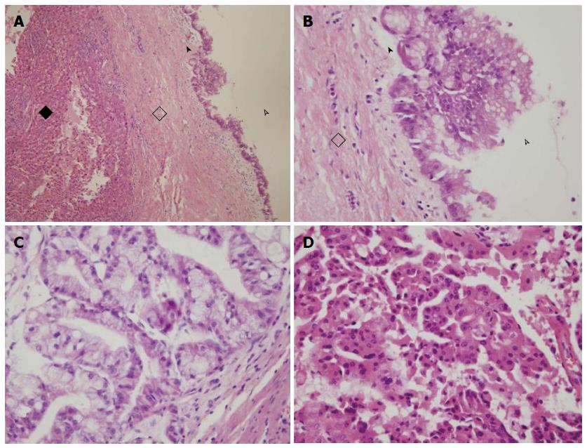
Figure 1 Pathology of intrahepatic biliary cystadenoma and cystadenocarcinoma.
A, B: Intrahepatic biliary cystadenoma (black diamond, hepatic tissue; hollow diamond, fibrous cyst wall; arrowhead, simple columnar epithelium; hollow arrowhead, cavity); C, D: Intrahepatic biliary cystadenocarcinoma. Mucinous cystadenocarcinoma with columnar epithelium, abundant cytoplasm, containing mucin, and nuclei located in the basal layer (C).
- Citation: Zhang FB, Zhang AM, Zhang ZB, Huang X, Wang XT, Dong JH. Preoperative differential diagnosis between intrahepatic biliary cystadenoma and cystadenocarcinoma: A single-center experience. World J Gastroenterol 2014; 20(35): 12595-12601
- URL: https://www.wjgnet.com/1007-9327/full/v20/i35/12595.htm
- DOI: https://dx.doi.org/10.3748/wjg.v20.i35.12595


