Copyright
©2014 Baishideng Publishing Group Inc.
World J Gastroenterol. Sep 21, 2014; 20(35): 12551-12558
Published online Sep 21, 2014. doi: 10.3748/wjg.v20.i35.12551
Published online Sep 21, 2014. doi: 10.3748/wjg.v20.i35.12551
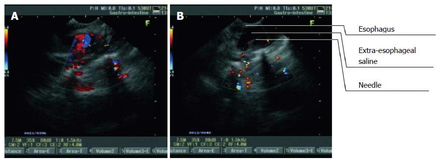
Figure 3 Procedure of extraesophageal saline injection guided by real-time linear-array endoscopic ultrasonography in the lower thoracic segment of the esophagus.
A: The thoracic aorta was visualized as turbulent blood flow on a color Doppler flow imaging (CDFI) image from L-EUS; B: The puncture probe was rotated until the thoracic aorta disappeared. Extraesophageal puncture, reverse suction, and saline injection were then performed consecutively. With an increase in the volume of injected saline, the hypoechoic area outside the esophagus expanded.
- Citation: Li JJ, Shan HB, He LJ, Wang TD, Xiong H, Chen LM, Li XH, Huang XX, Luo GY, Li Y, Xu GL. Extraesophageal saline enhances endoscopic ultrasonography to differentiate esophagus and adjacent organs. World J Gastroenterol 2014; 20(35): 12551-12558
- URL: https://www.wjgnet.com/1007-9327/full/v20/i35/12551.htm
- DOI: https://dx.doi.org/10.3748/wjg.v20.i35.12551


