Copyright
©2014 Baishideng Publishing Group Inc.
World J Gastroenterol. Sep 21, 2014; 20(35): 12445-12457
Published online Sep 21, 2014. doi: 10.3748/wjg.v20.i35.12445
Published online Sep 21, 2014. doi: 10.3748/wjg.v20.i35.12445
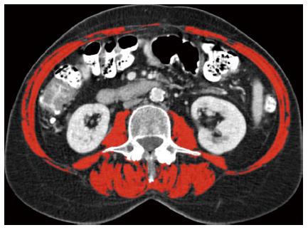
Figure 1 Computed tomography image at the third lumbar vertebral level.
The following skeletal muscles are outlined in red: psoas, paraspinal, transverse abdominal, external oblique, internal oblique and rectus abdominis muscles. This female sarcopenia patient had an L3 (third lumbar spine) muscle index of 34.3 cm²/m².
- Citation: van Vugt JL, Reisinger KW, Derikx JP, Boerma D, Stoot JH. Improving the outcomes in oncological colorectal surgery. World J Gastroenterol 2014; 20(35): 12445-12457
- URL: https://www.wjgnet.com/1007-9327/full/v20/i35/12445.htm
- DOI: https://dx.doi.org/10.3748/wjg.v20.i35.12445


