Copyright
©2014 Baishideng Publishing Group Inc.
World J Gastroenterol. Sep 21, 2014; 20(35): 12381-12390
Published online Sep 21, 2014. doi: 10.3748/wjg.v20.i35.12381
Published online Sep 21, 2014. doi: 10.3748/wjg.v20.i35.12381
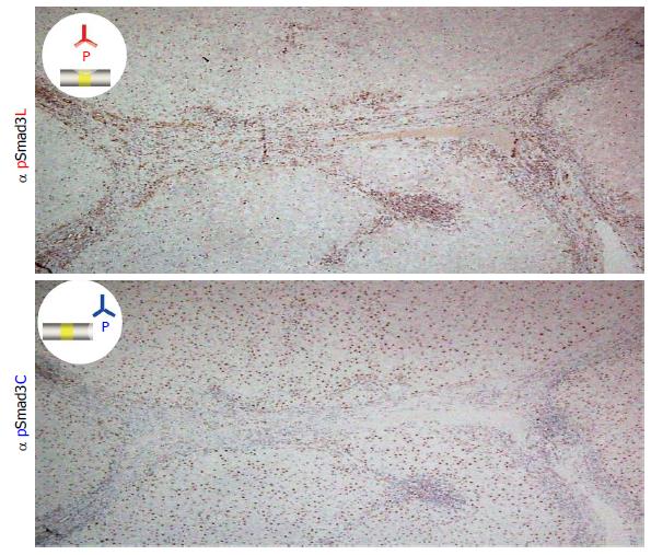
Figure 1 Two distinct hepatocytic Smad3 phospho-isoforms in chronic hepatitis C.
Liver specimens with moderate fibrosis and inflammation from a patient with chronic hepatitis C were immunostained using domain-specific antibodies (Abs) that are able to distinguish between the phosphorylated linker region and the C-terminal region of Smad3. Each section was counterstained with hematoxylin (blue). The brown color indicates specific Ab reactivity. The distribution of pSmad3C and pSmad3L showed a sharp contrast in the liver specimens. Whereas the hepatocytes adjacent to the collagen fibers of the portal tract showed nuclear localization of pSmad3L (upper column), pSmad3C was predominantly located in the hepatocytic nuclei distant from the portal tract (lower column).
- Citation: Yamaguchi T, Yoshida K, Murata M, Matsuzaki K. Smad3 phospho-isoform signaling in hepatitis C virus-related chronic liver diseases. World J Gastroenterol 2014; 20(35): 12381-12390
- URL: https://www.wjgnet.com/1007-9327/full/v20/i35/12381.htm
- DOI: https://dx.doi.org/10.3748/wjg.v20.i35.12381


