Copyright
©2014 Baishideng Publishing Group Inc.
World J Gastroenterol. Sep 14, 2014; 20(34): 12346-12349
Published online Sep 14, 2014. doi: 10.3748/wjg.v20.i34.12346
Published online Sep 14, 2014. doi: 10.3748/wjg.v20.i34.12346
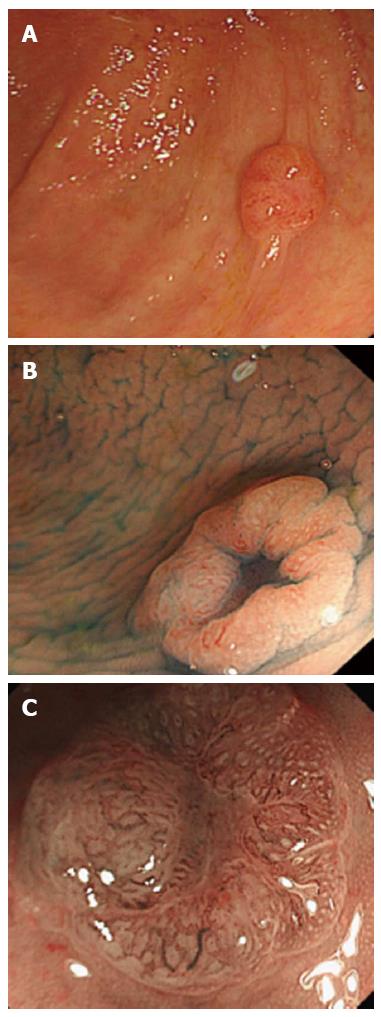
Figure 1 Endoscopic findings in our case.
A: Colonoscopic view showing a 7-mm flat-elevated lesion in the cecum; B: Chromocolonoscopic view (indigo carmine dye) showing a central depression in the lesion; C: Magnifying narrow-band imaging of the depressed area showing winding and prematurely terminating irregular blood vessels and sparse surface patterns.
- Citation: Aoki H, Nosho K, Igarashi H, Ito M, Mitsuhashi K, Naito T, Yamamoto E, Tanuma T, Nomura M, Maguchi H, Shinohara T, Suzuki H, Yamamoto H, Shinomura Y. MicroRNA-31 expression in colorectal serrated pathway progression. World J Gastroenterol 2014; 20(34): 12346-12349
- URL: https://www.wjgnet.com/1007-9327/full/v20/i34/12346.htm
- DOI: https://dx.doi.org/10.3748/wjg.v20.i34.12346


