Copyright
©2014 Baishideng Publishing Group Inc.
World J Gastroenterol. Sep 14, 2014; 20(34): 12341-12345
Published online Sep 14, 2014. doi: 10.3748/wjg.v20.i34.12341
Published online Sep 14, 2014. doi: 10.3748/wjg.v20.i34.12341
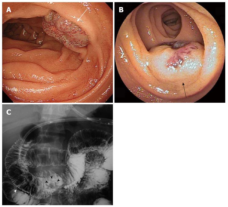
Figure 1 Hemangiomas in the duodenum and jejunum.
A: Upper gastrointestinal endoscopy shows a hemangioma of the duodenum in the third portion (white arrow); B: Double balloon endoscopy shows a hemangioma of the jejunum (black arrow); C: A barium study shows the duodenal hemangioma (black arrowheads) located in the third portion of duodenum, approximately 4.0 cm from the anal side of the inferior flexure (white arrowheads).
- Citation: Kanaji S, Nakamura T, Nishi M, Yamamoto M, Kanemitu K, Yamashiita K, Imanishi T, Sumi Y, Suzuki S, Tanaka K, Kakeji Y. Laparoscopic partial resection for hemangioma in the third portion of the duodenum. World J Gastroenterol 2014; 20(34): 12341-12345
- URL: https://www.wjgnet.com/1007-9327/full/v20/i34/12341.htm
- DOI: https://dx.doi.org/10.3748/wjg.v20.i34.12341


