Copyright
©2014 Baishideng Publishing Group Inc.
World J Gastroenterol. Sep 14, 2014; 20(34): 12171-12181
Published online Sep 14, 2014. doi: 10.3748/wjg.v20.i34.12171
Published online Sep 14, 2014. doi: 10.3748/wjg.v20.i34.12171
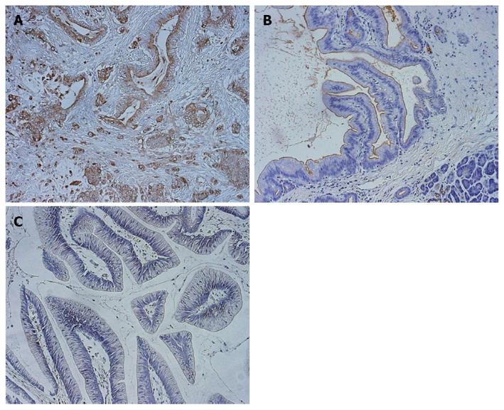
Figure 1 Immunohistochemistry for KL-6/MUC1 expression in tissue.
Staining for KL-6/MUC1 in A: Pancreatic duct cell carcinoma; B: Pancreatic duct cell carcinoma and surrounding normal tissues; C: Intraductal papillary mucinous tumor (200 × magnification).
- Citation: Xu HL, Zhao X, Zhang KM, Tang W, Kokudo N. Inhibition of KL-6/MUC1 glycosylation limits aggressive progression of pancreatic cancer. World J Gastroenterol 2014; 20(34): 12171-12181
- URL: https://www.wjgnet.com/1007-9327/full/v20/i34/12171.htm
- DOI: https://dx.doi.org/10.3748/wjg.v20.i34.12171


