Copyright
©2014 Baishideng Publishing Group Inc.
World J Gastroenterol. Sep 14, 2014; 20(34): 12118-12131
Published online Sep 14, 2014. doi: 10.3748/wjg.v20.i34.12118
Published online Sep 14, 2014. doi: 10.3748/wjg.v20.i34.12118
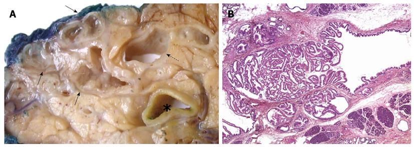
Figure 1 Intraductal papillary mucinous neoplasia.
A: The main pancreatic duct (dotted arrow) and a cluster of branch ducts (arrows) are dilated, and particularly the latter contain mucus and a papillary proliferation on the duct walls (asterisk, common bile duct); B: Partial involvement of a pancreatic branch duct by the papillary proliferation of neoplastic epithelium characteristic of intraductal papillary mucinous neoplasia (HE, 20 × magnification).
- Citation: Chiaro MD, Segersvärd R, Lohr M, Verbeke C. Early detection and prevention of pancreatic cancer: Is it really possible today? World J Gastroenterol 2014; 20(34): 12118-12131
- URL: https://www.wjgnet.com/1007-9327/full/v20/i34/12118.htm
- DOI: https://dx.doi.org/10.3748/wjg.v20.i34.12118


