Copyright
©2014 Baishideng Publishing Group Inc.
World J Gastroenterol. Sep 7, 2014; 20(33): 11921-11926
Published online Sep 7, 2014. doi: 10.3748/wjg.v20.i33.11921
Published online Sep 7, 2014. doi: 10.3748/wjg.v20.i33.11921
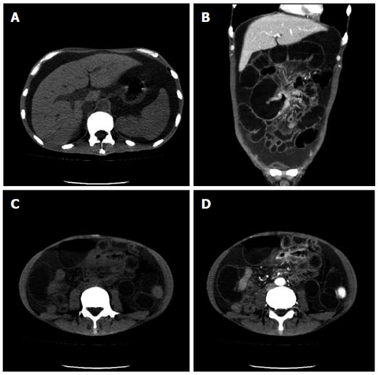
Figure 2 Computerized tomography images indicated hepatosplenomegaly and ascites (A) and the presence of thickened intestinal wall with obvious enhancement in the arterial phase and dilated small bowel (B-D).
A: Plain computerized tomography (CT) scan; B: Coronal CT scan; C: Plain CT scan; D: The arterial phase.
- Citation: Wei XQ, Zheng ZH, Jin Y, Tao J, Abassa KK, Wen ZF, Shao CK, Wei HB, Wu B. Intestinal obstruction caused by extramedullary hematopoiesis and ascites in primary myelofibrosis. World J Gastroenterol 2014; 20(33): 11921-11926
- URL: https://www.wjgnet.com/1007-9327/full/v20/i33/11921.htm
- DOI: https://dx.doi.org/10.3748/wjg.v20.i33.11921


