Copyright
©2014 Baishideng Publishing Group Inc.
World J Gastroenterol. Sep 7, 2014; 20(33): 11916-11920
Published online Sep 7, 2014. doi: 10.3748/wjg.v20.i33.11916
Published online Sep 7, 2014. doi: 10.3748/wjg.v20.i33.11916
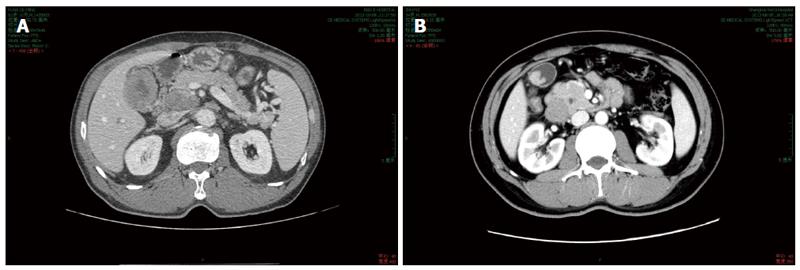
Figure 1 Computed tomography scan of the gallbladder carcinoma.
A: A thick-walled gallbladder with local infiltration to the adjacent liver; B: A polypoid intra-luminal lesion about 4 cm in diameter.
- Citation: Chen H, Shen YY, Ni XZ. Two cases of neuroendocrine carcinoma of the gallbladder. World J Gastroenterol 2014; 20(33): 11916-11920
- URL: https://www.wjgnet.com/1007-9327/full/v20/i33/11916.htm
- DOI: https://dx.doi.org/10.3748/wjg.v20.i33.11916


