Copyright
©2014 Baishideng Publishing Group Inc.
World J Gastroenterol. Sep 7, 2014; 20(33): 11904-11909
Published online Sep 7, 2014. doi: 10.3748/wjg.v20.i33.11904
Published online Sep 7, 2014. doi: 10.3748/wjg.v20.i33.11904
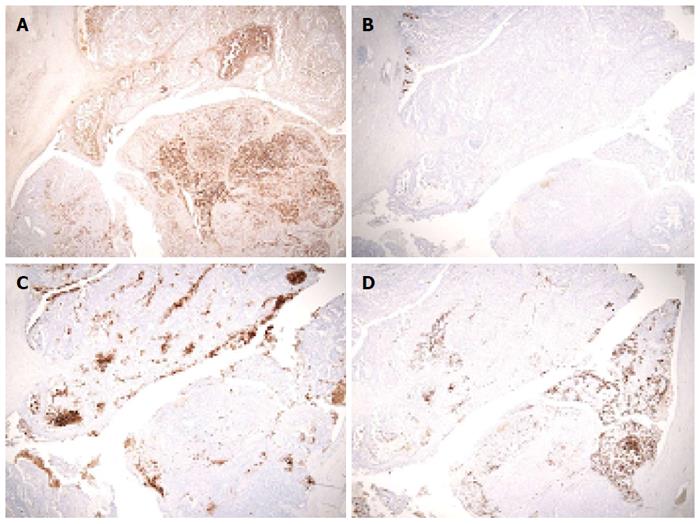
Figure 6 Immunohistochemical and pathological findings from the second operation.
A: α1-antitrypsin (× 40): the acinar structure in the lower right area was strongly positive; B: Mucin5AC (× 40): only a small part of the tumor was positively stained; C: Carcinoembryonic antigen (× 40): only a portion of the area was positively stained; D: Mucin1 (× 40): some positive staining was observed.
- Citation: Shonaka T, Inagaki M, Akabane H, Yanagida N, Shomura H, Yanagawa N, Oikawa K, Nakano S. Total pancreatectomy for metachronous mixed acinar-ductal carcinoma in a remnant pancreas. World J Gastroenterol 2014; 20(33): 11904-11909
- URL: https://www.wjgnet.com/1007-9327/full/v20/i33/11904.htm
- DOI: https://dx.doi.org/10.3748/wjg.v20.i33.11904


