Copyright
©2014 Baishideng Publishing Group Inc.
World J Gastroenterol. Sep 7, 2014; 20(33): 11904-11909
Published online Sep 7, 2014. doi: 10.3748/wjg.v20.i33.11904
Published online Sep 7, 2014. doi: 10.3748/wjg.v20.i33.11904
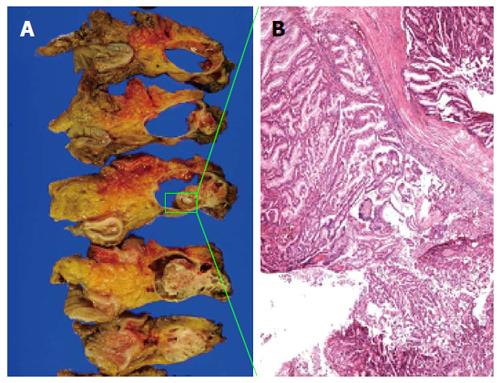
Figure 3 The resected specimen and pathological findings from the first operation.
A: The resected specimen had a cystic lesion with a solid component; B: HE staining showed a mucus-type epithelium with an extension of intraductal papillary growth of the tumor cells. Scarce mucus-producing cells were also present (HE, × 40).
- Citation: Shonaka T, Inagaki M, Akabane H, Yanagida N, Shomura H, Yanagawa N, Oikawa K, Nakano S. Total pancreatectomy for metachronous mixed acinar-ductal carcinoma in a remnant pancreas. World J Gastroenterol 2014; 20(33): 11904-11909
- URL: https://www.wjgnet.com/1007-9327/full/v20/i33/11904.htm
- DOI: https://dx.doi.org/10.3748/wjg.v20.i33.11904


