Copyright
©2014 Baishideng Publishing Group Inc.
World J Gastroenterol. Sep 7, 2014; 20(33): 11840-11849
Published online Sep 7, 2014. doi: 10.3748/wjg.v20.i33.11840
Published online Sep 7, 2014. doi: 10.3748/wjg.v20.i33.11840
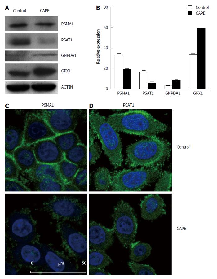
Figure 3 Validation of differentially expressed proteins in SW480 treated with caffeic acid phenethyl ester.
SW480 cells were treated without (control) and with 10 μg/mL CAPE for 48 h and then harvested for Western blotting (A and B) or immunofluorescence assay (C and D). For Western blotting, beta-actin was included as the internal control. Densitometric analysis was performed and the integrated density values are presented as the ratio of each protein over the beta-actin protein (B). For the immunofluorescence assay, SW480 cells were grown on glass coverslips and treated without (control) or with CAPE (10 μg/mL) for 48 h. The cells were washed with PBS and fixed with methanol. Anti-PSMA1 (A), and anti-PSAT1 (B) monoclonal antibodies were applied as the primary antibodies, and then, FITC-conjugated secondary antibodies were used. DAPI was used to stain the nucleus. Results are representative of 2 independent experiments with similar results. PSMA1: Proteasome subunit alpha 1; PSAT1: Phosphoserine aminotransferase 1.
- Citation: He YJ, Li WL, Liu BH, Dong H, Mou ZR, Wu YZ. Identification of differential proteins in colorectal cancer cells treated with caffeic acid phenethyl ester. World J Gastroenterol 2014; 20(33): 11840-11849
- URL: https://www.wjgnet.com/1007-9327/full/v20/i33/11840.htm
- DOI: https://dx.doi.org/10.3748/wjg.v20.i33.11840


