Copyright
©2014 Baishideng Publishing Group Inc.
World J Gastroenterol. Sep 7, 2014; 20(33): 11650-11670
Published online Sep 7, 2014. doi: 10.3748/wjg.v20.i33.11650
Published online Sep 7, 2014. doi: 10.3748/wjg.v20.i33.11650
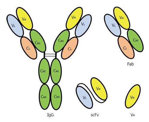
Figure 4 Recombinant antibodies displayed on bacteriophage compared with an immunoglobulin G molecule.
Chain structure of a human immunoglobulin G (IgG) molecule. V: Variable region; C: Constant region; H: Heavy chain; L: Light chain; VH: Variable region of heavy chain; Fab: Fragment antigen binding; scFv: Single chain fragment variable, the VL and VH are linked by a peptide linker.
- Citation: Tan WS, Ho KL. Phage display creates innovative applications to combat hepatitis B virus. World J Gastroenterol 2014; 20(33): 11650-11670
- URL: https://www.wjgnet.com/1007-9327/full/v20/i33/11650.htm
- DOI: https://dx.doi.org/10.3748/wjg.v20.i33.11650


