Copyright
©2014 Baishideng Publishing Group Inc.
World J Gastroenterol. Aug 28, 2014; 20(32): 11451-11455
Published online Aug 28, 2014. doi: 10.3748/wjg.v20.i32.11451
Published online Aug 28, 2014. doi: 10.3748/wjg.v20.i32.11451
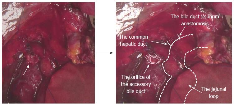
Figure 5 Intraoperative findings in the second operation.
The orifice of the injured caudate lobe bile duct of the proximal side on the surface of the liver was recognised (arrow). The precise anatomy is described using the annotation method.
- Citation: Miyamoto R, Oshiro Y, Hashimoto S, Kohno K, Fukunaga K, Oda T, Ohkohchi N. Three-dimensional imaging identified the accessory bile duct in a patient with cholangiocarcinoma. World J Gastroenterol 2014; 20(32): 11451-11455
- URL: https://www.wjgnet.com/1007-9327/full/v20/i32/11451.htm
- DOI: https://dx.doi.org/10.3748/wjg.v20.i32.11451


