Copyright
©2014 Baishideng Publishing Group Inc.
World J Gastroenterol. Aug 28, 2014; 20(32): 11451-11455
Published online Aug 28, 2014. doi: 10.3748/wjg.v20.i32.11451
Published online Aug 28, 2014. doi: 10.3748/wjg.v20.i32.11451
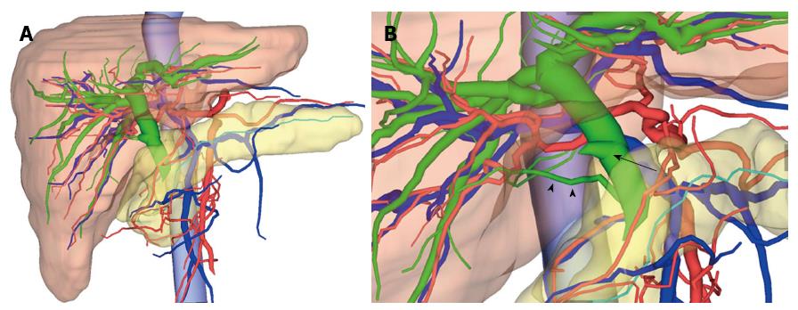
Figure 4 3-dimensional images by integrating multidetector computed tomography and magnetic resonance cholangiopancreatography images.
A: 3-dimensional (3D) image view from the front side of the patient. The red colour represents the arteries, the blue represents the veins and the portal vein, the green represents the biliary duct, and the turquoise represents the pancreatic duct. We were able to observe that the common and intrahepatic bile ducts were dilated; B: From the 3D image view from the patient's right side, the accessory bile duct from the caudate lobe connecting to the intrapancreatic bile duct (arrowheads) was easily recognisable. The cystic duct (arrow) has branched from the middle bile duct. We were able to determine that the injured site was the accessory bile duct from the caudate lobe.
- Citation: Miyamoto R, Oshiro Y, Hashimoto S, Kohno K, Fukunaga K, Oda T, Ohkohchi N. Three-dimensional imaging identified the accessory bile duct in a patient with cholangiocarcinoma. World J Gastroenterol 2014; 20(32): 11451-11455
- URL: https://www.wjgnet.com/1007-9327/full/v20/i32/11451.htm
- DOI: https://dx.doi.org/10.3748/wjg.v20.i32.11451


