Copyright
©2014 Baishideng Publishing Group Inc.
World J Gastroenterol. Aug 28, 2014; 20(32): 11451-11455
Published online Aug 28, 2014. doi: 10.3748/wjg.v20.i32.11451
Published online Aug 28, 2014. doi: 10.3748/wjg.v20.i32.11451
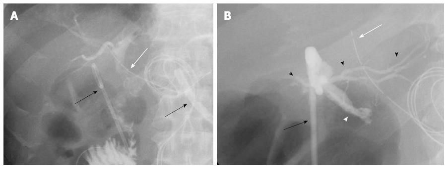
Figure 3 Tubography through the biliary drainage tube (white arrow) and the abdominal drainage tube (black arrow) after the first operation.
A: The radiological image indicated the intrahepatic bile duct, common bile duct, and jejunum. Leakage of the contrast media from the end to side biliojejunostomy was not observed. Two abdominal drainage tubes (black arrows) were inserted into the abdominal cavity; B: Postoperative tubography through the abdominal drainage tube (black arrow) was performed, which produced the images of the injured site of the probable caudate bile duct (white arrowhead) and the other intrahepatic bile duct (black arrowheads). The biliary drainage tube is shown (white arrow).
- Citation: Miyamoto R, Oshiro Y, Hashimoto S, Kohno K, Fukunaga K, Oda T, Ohkohchi N. Three-dimensional imaging identified the accessory bile duct in a patient with cholangiocarcinoma. World J Gastroenterol 2014; 20(32): 11451-11455
- URL: https://www.wjgnet.com/1007-9327/full/v20/i32/11451.htm
- DOI: https://dx.doi.org/10.3748/wjg.v20.i32.11451


