Copyright
©2014 Baishideng Publishing Group Inc.
World J Gastroenterol. Aug 28, 2014; 20(32): 11305-11312
Published online Aug 28, 2014. doi: 10.3748/wjg.v20.i32.11305
Published online Aug 28, 2014. doi: 10.3748/wjg.v20.i32.11305
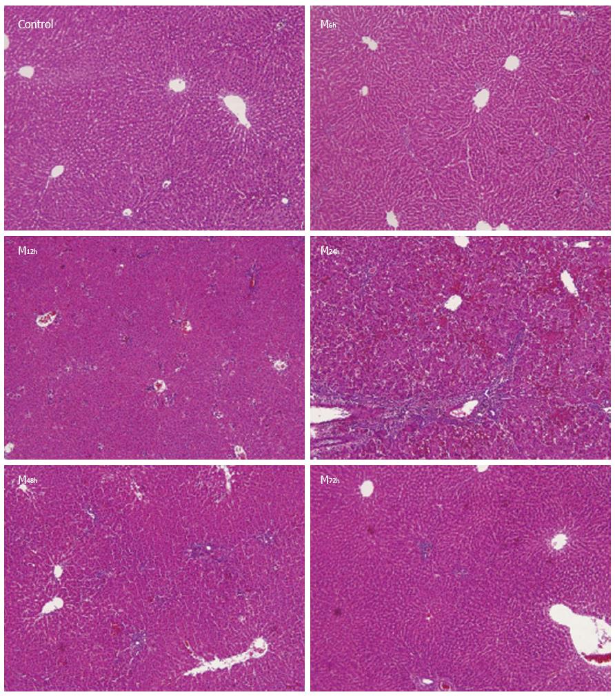
Figure 1 Histopathological findings of liver tissues in control and model groups (hematoxylin and eosin × 100).
Control: Hepatic architecture was normal with little necrosis and inflammatory cell infiltration; M6h: The structure of the hepatic lobules was largely normal with spotty, focal necrosis and little inflammatory cell infiltration; M12h: The structure of hepatic lobules was slightly damaged, with focal necrosis and little inflammatory cell infiltration; M24h: There was an extensive area of central-central and central-portal bridging necrosis containing many red cells trapped in the collapsed reticulin network, and significant inflammatory cell infiltration was observed; M48h: Hepatic necrosis and inflammatory cell infiltration gradually reduced; M72h: Hepatic lobular structure returned to normal, with very little inflammatory cell infiltration.
- Citation: Zhu D, Zhang H, Mao JY, Wang HY, Li X, Xu YQ. Role of the 13C-methacetin breath test in the assessment of acute liver injury in a rat model. World J Gastroenterol 2014; 20(32): 11305-11312
- URL: https://www.wjgnet.com/1007-9327/full/v20/i32/11305.htm
- DOI: https://dx.doi.org/10.3748/wjg.v20.i32.11305


