Copyright
©2014 Baishideng Publishing Group Inc.
World J Gastroenterol. Aug 28, 2014; 20(32): 11131-11141
Published online Aug 28, 2014. doi: 10.3748/wjg.v20.i32.11131
Published online Aug 28, 2014. doi: 10.3748/wjg.v20.i32.11131
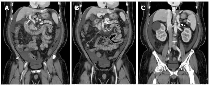
Figure 5 Preoperative computed tomographic findings of case 2.
A: The right lobe portal vein could not be identified, likely owing to hepatofugal flow-induced stasis and thrombosis (white arrowhead) and portal-systemic shunting from the left portal vein to the left pericardiac-phrenic vein (white asterisk); B: Coronary vein engorgement (double white asterisks); C: Spontaneous splenorenal shunting was also noted (white arrow).
- Citation: Feng AC, Fan HL, Chen TW, Hsieh CB. Hepatic hemodynamic changes during liver transplantation: A review. World J Gastroenterol 2014; 20(32): 11131-11141
- URL: https://www.wjgnet.com/1007-9327/full/v20/i32/11131.htm
- DOI: https://dx.doi.org/10.3748/wjg.v20.i32.11131


