Copyright
©2014 Baishideng Publishing Group Inc.
World J Gastroenterol. Aug 28, 2014; 20(32): 11080-11094
Published online Aug 28, 2014. doi: 10.3748/wjg.v20.i32.11080
Published online Aug 28, 2014. doi: 10.3748/wjg.v20.i32.11080
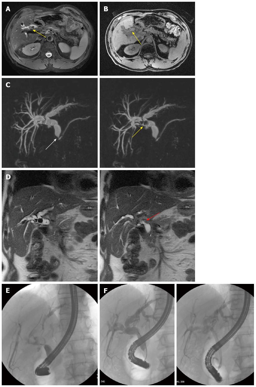
Figure 5 Anastomotic biliary stricture with lithiasis.
A: Axial T2-weighted image shows dilation of the biliary system with concomitant stones (yellow arrow); B: Axial T1-weighted image confirms the presence of stones in the biliary tract (yellow arrow); C: Maximum intensity projections of 3D thin-slab fast spin-echo T2-weighted images (obtained using different thicknesses) demonstrate the dilation of the both intra- and extra-hepatic (pre- and post-anastomotic) biliary tract with a stricture of the iuxta-papillary choledocho (white arrow); the presence of two stones at the level of the hepatic bifurcation (yellow arrow) is also well appreciable; D: On coronal single-shot T2-weighted images (at different levels) is also better appreciable a stricture at the anastomotic site (red arrow); E: Endoscopic retrograde cholangiography confirms the presence of strictures and stones in the pre-anastomotic biliary tract; F: Stones were endoscopically removed and strictures were treated by stenting as shown on different projection images.
- Citation: Boraschi P, Donati F. Postoperative biliary adverse events following orthotopic liver transplantation: Assessment with magnetic resonance cholangiography. World J Gastroenterol 2014; 20(32): 11080-11094
- URL: https://www.wjgnet.com/1007-9327/full/v20/i32/11080.htm
- DOI: https://dx.doi.org/10.3748/wjg.v20.i32.11080


