Copyright
©2014 Baishideng Publishing Group Inc.
World J Gastroenterol. Aug 28, 2014; 20(32): 11080-11094
Published online Aug 28, 2014. doi: 10.3748/wjg.v20.i32.11080
Published online Aug 28, 2014. doi: 10.3748/wjg.v20.i32.11080
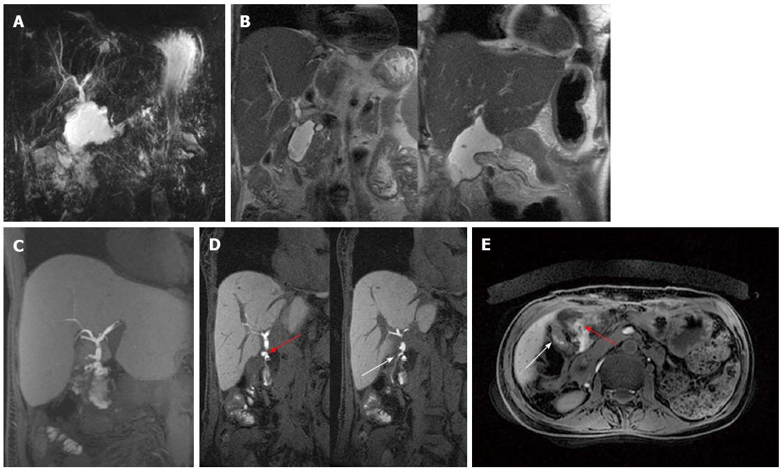
Figure 3 Anastomotic leak in a patient with hepatico-jejunostomy.
A: Single-shot thick-slab magnetic resonance cholangiogram shows a fluid collection in the area of biliary-enteric anastomosis; B: Coronal T2-weighted images (at different levels) accurately depict circumscribed sub-hepatic fluid collections with thickened walls. C: Maximum intensity projection reconstruction of Gd-EOB-DTPA enhanced LAVA T1-weighted sequence well exhibits extravasation of contrast material into the peri-anastomotic space compatible with bile leak; D: Coronal Gd-EOB-DTPA enhanced LAVA T1-weighted images better identify contrast agent both extravasating from an anastomotic leak (red arrow) and filling the Roux-en-Y anastomosis (white arrow); E: On axial post-contrast LAVA image it is possible to distinguish the fluid collection (red arrow) from the jejunum (white arrow).
- Citation: Boraschi P, Donati F. Postoperative biliary adverse events following orthotopic liver transplantation: Assessment with magnetic resonance cholangiography. World J Gastroenterol 2014; 20(32): 11080-11094
- URL: https://www.wjgnet.com/1007-9327/full/v20/i32/11080.htm
- DOI: https://dx.doi.org/10.3748/wjg.v20.i32.11080


