Copyright
©2014 Baishideng Publishing Group Inc.
World J Gastroenterol. Aug 28, 2014; 20(32): 11080-11094
Published online Aug 28, 2014. doi: 10.3748/wjg.v20.i32.11080
Published online Aug 28, 2014. doi: 10.3748/wjg.v20.i32.11080
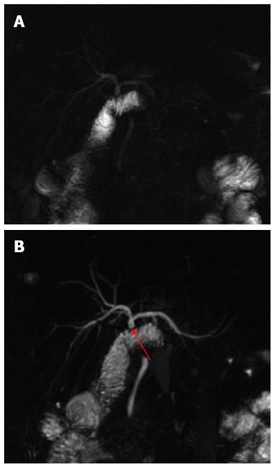
Figure 1 Bilio-enteric anastomosis.
A: Single-shot thick-slab magnetic resonance cholangiogram shows a regular hepatico-jejunostomy; B: Maximum intensity projection reconstruction of 3D thin-slab fast spin-echo T2-weighted images confirms the anastomotic patency (red arrow) and better demonstrates the portion of the jejunum and the choledocho of the recipient.
- Citation: Boraschi P, Donati F. Postoperative biliary adverse events following orthotopic liver transplantation: Assessment with magnetic resonance cholangiography. World J Gastroenterol 2014; 20(32): 11080-11094
- URL: https://www.wjgnet.com/1007-9327/full/v20/i32/11080.htm
- DOI: https://dx.doi.org/10.3748/wjg.v20.i32.11080


