Copyright
©2014 Baishideng Publishing Group Inc.
World J Gastroenterol. Aug 21, 2014; 20(31): 10864-10875
Published online Aug 21, 2014. doi: 10.3748/wjg.v20.i31.10864
Published online Aug 21, 2014. doi: 10.3748/wjg.v20.i31.10864
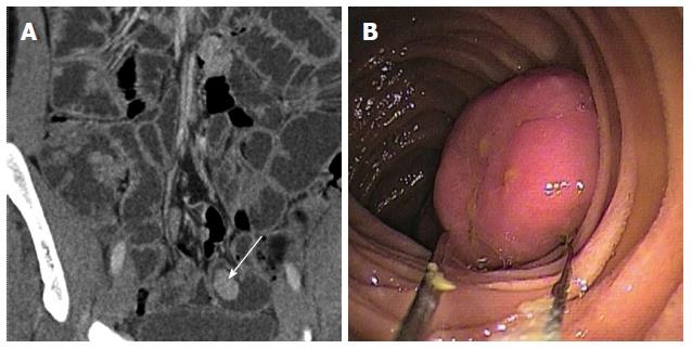
Figure 4 Polyp 4.
A: Multidetector computed tomography (MDCT) coronal view reveals a regular small-bowel polyp with homogeneous enhancement (arrow); B: The double balloon endoscopy optimally depicts this large small bowel polyp.
- Citation: Tomas C, Soyer P, Dohan A, Dray X, Boudiaf M, Hoeffel C. Update on imaging of Peutz-Jeghers syndrome. World J Gastroenterol 2014; 20(31): 10864-10875
- URL: https://www.wjgnet.com/1007-9327/full/v20/i31/10864.htm
- DOI: https://dx.doi.org/10.3748/wjg.v20.i31.10864


