Copyright
©2014 Baishideng Publishing Group Inc.
World J Gastroenterol. Aug 14, 2014; 20(30): 10642-10650
Published online Aug 14, 2014. doi: 10.3748/wjg.v20.i30.10642
Published online Aug 14, 2014. doi: 10.3748/wjg.v20.i30.10642
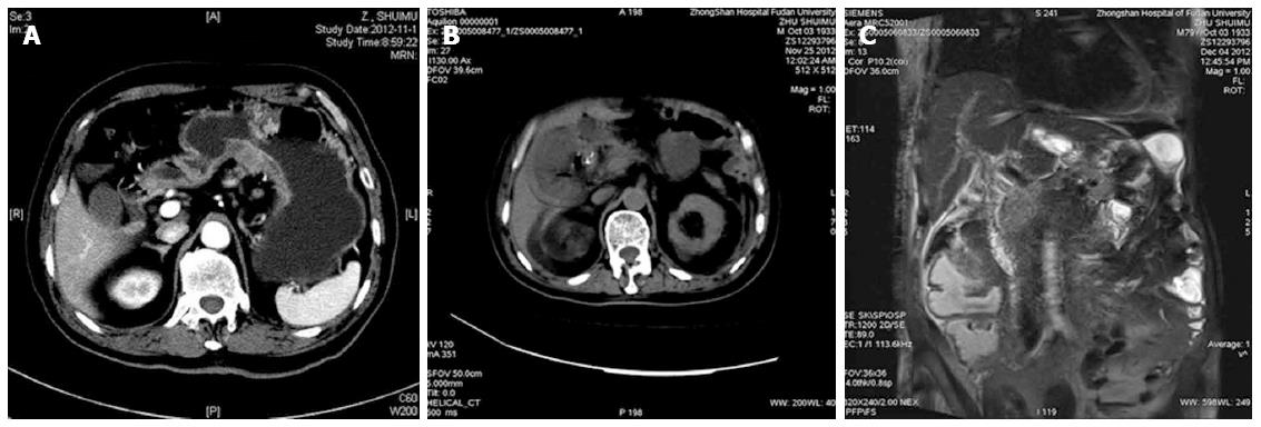
Figure 2 Computed tomography scan in case 2 displaying normal gallbladder (A) before operation, thickened gallbladder wall and swelling gallbladder (B), and magnetic resonance imaging showing no dilation of bile ducts on the 19th day after operation (C).
- Citation: Liu FL, Li H, Wang XF, Shen KT, Shen ZB, Sun YH, Qin XY. Acute acalculous cholecystitis immediately after gastric operation: Case report and literatures review. World J Gastroenterol 2014; 20(30): 10642-10650
- URL: https://www.wjgnet.com/1007-9327/full/v20/i30/10642.htm
- DOI: https://dx.doi.org/10.3748/wjg.v20.i30.10642


