Copyright
©2014 Baishideng Publishing Group Inc.
World J Gastroenterol. Aug 14, 2014; 20(30): 10470-10477
Published online Aug 14, 2014. doi: 10.3748/wjg.v20.i30.10470
Published online Aug 14, 2014. doi: 10.3748/wjg.v20.i30.10470
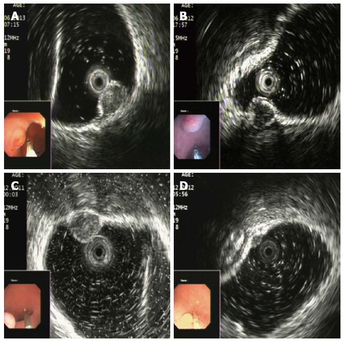
Figure 2 Endoscopic ultrasonography findings of rectal neuroendocrine neoplasms.
A: An egg-shaped lesion within the mucosa with an intermediate echo pattern and distinct border; B: A homogenous hypoechoic lesion with a distinct border within the submucosa; C: A round medium-echo lesion with a distinct border within the submucosa; D: A nodular neuroendocrine neoplasm located in both the mucosa and submucosa.
- Citation: Chen HT, Xu GQ, Teng XD, Chen YP, Chen LH, Li YM. Diagnostic accuracy of endoscopic ultrasonography for rectal neuroendocrine neoplasms. World J Gastroenterol 2014; 20(30): 10470-10477
- URL: https://www.wjgnet.com/1007-9327/full/v20/i30/10470.htm
- DOI: https://dx.doi.org/10.3748/wjg.v20.i30.10470


