Copyright
©2014 Baishideng Publishing Group Co.
World J Gastroenterol. Jan 21, 2014; 20(3): 738-744
Published online Jan 21, 2014. doi: 10.3748/wjg.v20.i3.738
Published online Jan 21, 2014. doi: 10.3748/wjg.v20.i3.738
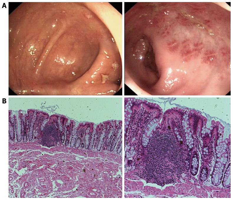
Figure 1 Endoscopic imaging and corresponding histological findings in solitary rectal ulcer syndrome patients.
A: Colonoscopy revealed localized yellowish slough, rectal edema, erythema, and superficial ulcerations; B: Histology (hematoxylin and eosin) shows smooth muscle hyperplasia in the lamina propria between colonic glands, and surface ulceration with associated chronic inflammatory infiltrates. Magnification: × 40 (left), × 100 (right).
- Citation: Zhu QC, Shen RR, Qin HL, Wang Y. Solitary rectal ulcer syndrome: Clinical features, pathophysiology, diagnosis and treatment strategies. World J Gastroenterol 2014; 20(3): 738-744
- URL: https://www.wjgnet.com/1007-9327/full/v20/i3/738.htm
- DOI: https://dx.doi.org/10.3748/wjg.v20.i3.738


