Copyright
©2014 Baishideng Publishing Group Inc.
World J Gastroenterol. Aug 7, 2014; 20(29): 10202-10207
Published online Aug 7, 2014. doi: 10.3748/wjg.v20.i29.10202
Published online Aug 7, 2014. doi: 10.3748/wjg.v20.i29.10202
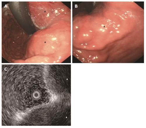
Figure 3 Endoscopy.
Endoscopy revealed irregular bulging (arrowhead) from the fundus to the posterior side of the midbody (A and B), and endosonography indicated effacement of the normal structure and a low echogenic mass (arrowhead) in the gastric wall (C).
- Citation: Zhao ZH, Yang JF, Wang JD, Wei JG, Liu F, Wang BY. Imaging findings of primary gastric plasmacytoma: A case report. World J Gastroenterol 2014; 20(29): 10202-10207
- URL: https://www.wjgnet.com/1007-9327/full/v20/i29/10202.htm
- DOI: https://dx.doi.org/10.3748/wjg.v20.i29.10202


