Copyright
©2014 Baishideng Publishing Group Inc.
World J Gastroenterol. Aug 7, 2014; 20(29): 10071-10081
Published online Aug 7, 2014. doi: 10.3748/wjg.v20.i29.10071
Published online Aug 7, 2014. doi: 10.3748/wjg.v20.i29.10071
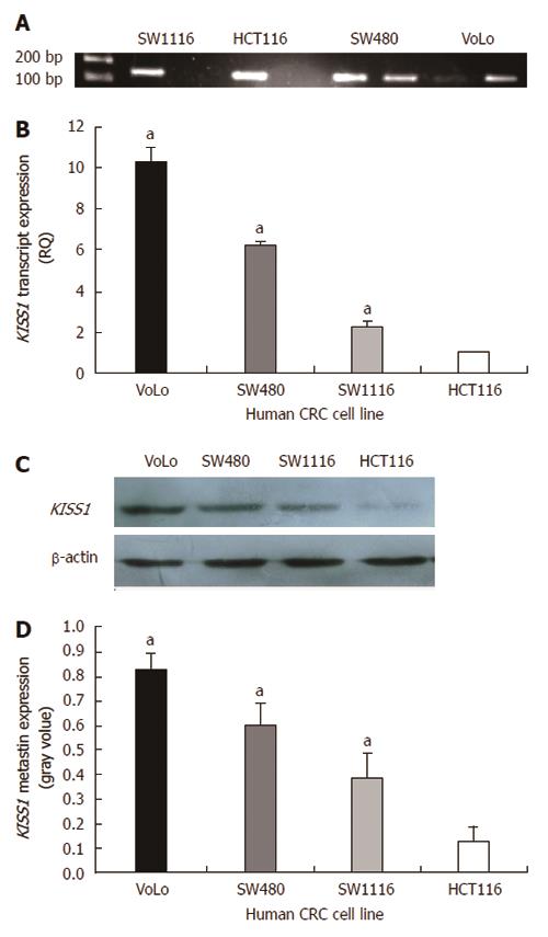
Figure 3 KISS1 methylation and KISS1 expression in various human colorectal cancer cell lines.
A: MS-PCR for KISS1 in human colorectal cancer (CRC) cell lines. A PCR band in lane M indicates a methylated KISS1 gene, whereas a band in lane U indicates an unmethylated KISS1 gene; B: KISS1 gene expression in CRC cell lines was examined by real-time PCR, and the expression of KISS1 was normalized to that of β-actin. The ratio of KISS1 expression in other CRC cells to KISS1 expression in HCT116 cells is shown on the Y-axis; C: Metastin expression in CRC cells of the indicated genotypes was determined by Western blot. The same membranes were subsequently probed with an anti-β-actin antibody as a loading control; D: The metastin expression levels are presented as the mean ± SEM. aP < 0.05 vs HCT116 cells.
-
Citation: Chen SQ, Chen ZH, Lin SY, Dai QB, Fu LX, Chen RQ.
KISS1 methylation and expression as predictors of disease progression in colorectal cancer patients. World J Gastroenterol 2014; 20(29): 10071-10081 - URL: https://www.wjgnet.com/1007-9327/full/v20/i29/10071.htm
- DOI: https://dx.doi.org/10.3748/wjg.v20.i29.10071


