Copyright
©2014 Baishideng Publishing Group Inc.
World J Gastroenterol. Jul 28, 2014; 20(28): 9618-9620
Published online Jul 28, 2014. doi: 10.3748/wjg.v20.i28.9618
Published online Jul 28, 2014. doi: 10.3748/wjg.v20.i28.9618
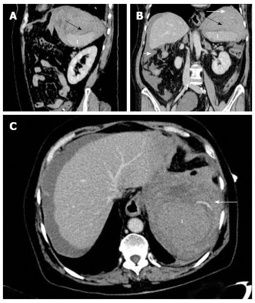
Figure 2 Computerized tomography images.
A, B: Multiplanar sagital and coronal reformation images: periesplenic hematoma (black arrow) and large hemoperitoneum (white arrows); C: Active splenic vascular contrast extravasation: focal areas of high attenuation (arrow) in splenic subcapsular space. 1Spleen.
- Citation: Tejada AH, Giménez-Alvira L, Brule EVD, Sánchez-Yuste R, Matallanos P, Blázquez E, Calleja JL, Abreu LE. Severe splenic rupture after colorectal endoscopic submucosal dissection. World J Gastroenterol 2014; 20(28): 9618-9620
- URL: https://www.wjgnet.com/1007-9327/full/v20/i28/9618.htm
- DOI: https://dx.doi.org/10.3748/wjg.v20.i28.9618


