Copyright
©2014 Baishideng Publishing Group Inc.
World J Gastroenterol. Jul 28, 2014; 20(28): 9564-9569
Published online Jul 28, 2014. doi: 10.3748/wjg.v20.i28.9564
Published online Jul 28, 2014. doi: 10.3748/wjg.v20.i28.9564
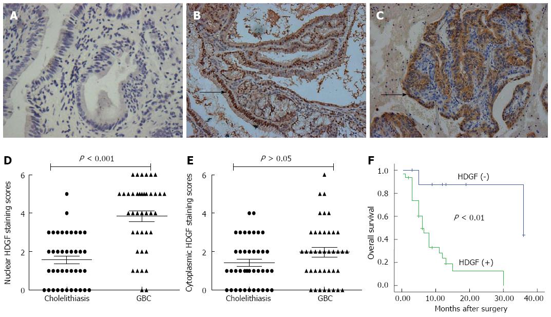
Figure 1 Enhanced expression of hepatoma-derived growth factor in gallbladder cancer and its negative association with prognosis.
A: Negative staining in normal glandular epithelium (× 200); B: Moderate staining predominantly localized in the cell nuclei in an adenocarcinoma. Arrows denote areas of positivity (× 200); C: Moderate to strong staining predominantly localized in the cell cytoplasm in an adenocarcinoma. Arrows denote areas of positivity (× 200); D: Average staining scores of nuclear hepatoma-derived growth factor (HDGF) expression in cholelithiasis and gallbladder cancer (GBC) tissues; E: Average staining scores of cytoplasmic HDGF expression in cholelithiasis and GBC tissues; F: Kaplan-Meier plots of overall survival in GBC patients with positive and negative HDGF expression scores.
- Citation: Tao F, Ye MF, Sun AJ, Lv JQ, Xu GG, Jing YM, Wang W. Prognostic significance of nuclear hepatoma-derived growth factor expression in gallbladder cancer. World J Gastroenterol 2014; 20(28): 9564-9569
- URL: https://www.wjgnet.com/1007-9327/full/v20/i28/9564.htm
- DOI: https://dx.doi.org/10.3748/wjg.v20.i28.9564


